
3-D Printing of Baby’s Heart Guides Surgery
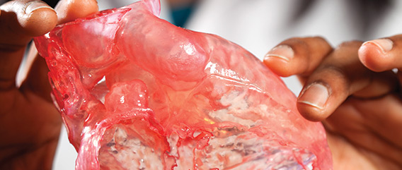
Columbia’s pediatric surgeons are now using 3-D printed models of patients’ hearts to guide surgery in children born with complex heart disease. The first model was used last October to treat a newborn with a rare and complex form of congenital heart disease, giving the surgeons an unrivalled understanding of the child’s defects and how to repair them in one operation.
“The baby’s heart had lots of holes, not uncommon with congenital disease, but the heart chambers were also in an unusual formation, like a maze,” says Emile Bacha, MD, the Calvin F. Barber Professor of Surgery at P&S and director of congenital and pediatric cardiac surgery at NewYork-Presbyterian/Morgan Stanley Children’s Hospital, where the surgery was performed.
“In the past, we had to stop the heart and look inside to decide what to do. But we only have a limited amount of time to perform the surgery,” he says. “With 3-D printing technology, we are able to look at the inside of the heart in advance, giving us a road map for the surgery.”
The first patient was just 1 week old when surgery was performed and had a heart the size of a walnut. With the aid of the 3-D model, printed to scale, the team was able to repair all of the heart’s defects in a single procedure. Typically, babies born with complex defects require three or four life-threatening surgeries.
Before the surgery, a team of doctors led by Anjali Chelliah, MD, assistant professor of pediatrics, diagnosed the baby with congenital heart disease while he was still in the womb, allowing time to develop the optimal treatment plan. One day after the baby was born, a low-dose CT scan and an echocardiogram provided excellent images, but given the size of the heart and the complexity of the defects, it was still difficult for the surgeons to see how the defects could be repaired.
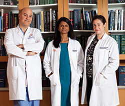 Emile Bacha, MD, Anjali Chelliah, MD, and Hannah Fraint, MD
Dr. Chelliah and pediatric cardiology fellow Hannah Fraint, MD, sent the CT imaging data to Materialise, a company that specializes in 3-D printing for health care, to create a model of the child’s heart. After seeing the model, the surgeons requested a different model cut at a different angle to gain even more understanding. They then devised a plan to fix all the defects in one seven-hour operation instead of three separate ones.
The patient is expected to not only have a normal lifespan, but also will probably not require any additional cardiac surgical interventions.
Dr. Bacha and Dr. Chelliah are optimistic that 3-D printing technology will continue to improve outcomes for patients. Since their first cases, they have printed 3-D model hearts of two preschool-age children; surgeries for both patients are being planned.
The physicians also are collecting the 3-D models in a library to help surgeons and cardiologists in training. “It’s clear that 3-D models can be successfully used to help surgeons in complex procedures,” says Dr. Bacha. “This technology is the future, and we are proud to help lead the way.”
The models used in this case were provided with the help of Matthew’s Hearts of Hope, a nonprofit organization, and a grant from the organization to Dr. Fraint.
- Log in to post comments